This guide will point you to appropriate groups based on the morphotype (amoeboid or spherical) and details of the pseudopodia.
Amoeboid Morphotype
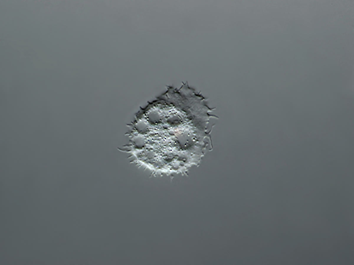
Without any differentiated locomotive organelles and with variable body shape; without a rigid pellicle.
Without any differentiated locomotive organelles and with variable body shape; without a rigid pellicle.
Pseudopodia: plasma projections with feeding, and/or sensing, and/or locomotion function.
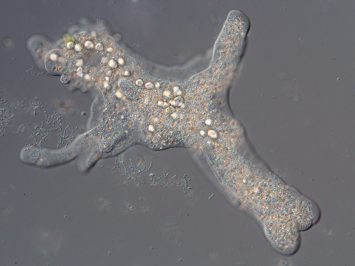
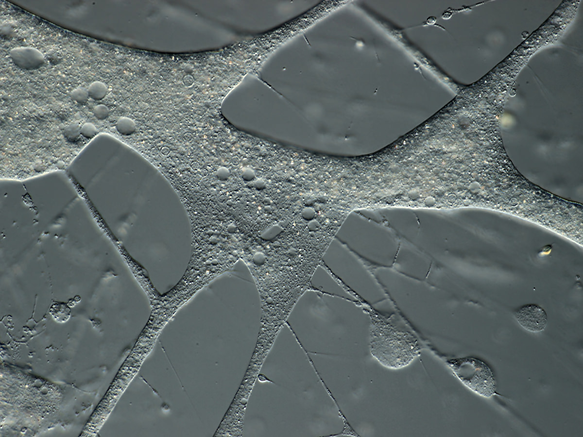
- Lobopodia: more or less broad, with cytoplasmic streaming; they may have a clear hyaline region at the front.
- Subpseudopodia: hyaline projections of different shape, usually anteriorly directed, which do not take part in the relocation of the main cell mass:
Dactylopodia: finger-shaped, hyaline pseudopodia.
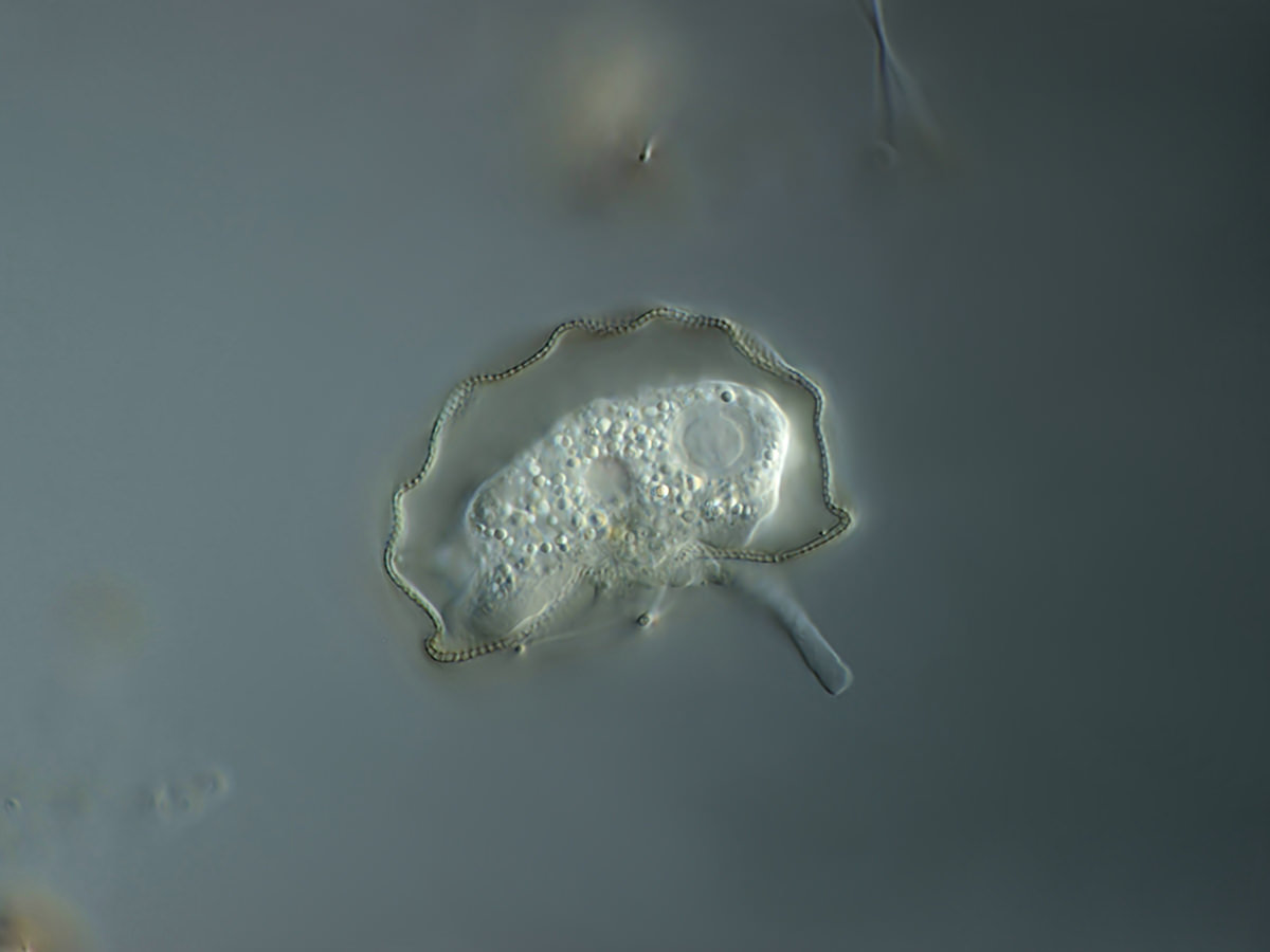
Lamellipodia: very thin and flat lobopodia; sometimes with filose tips at the margin.
dummy image - correct one will be added soon
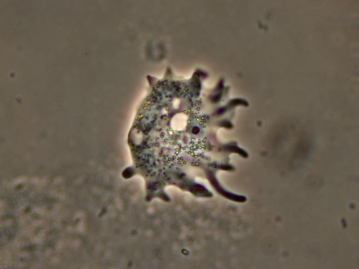
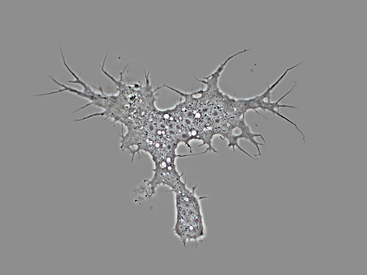
Acanthopodia: pointed, tooth-shaped pseudopodia.
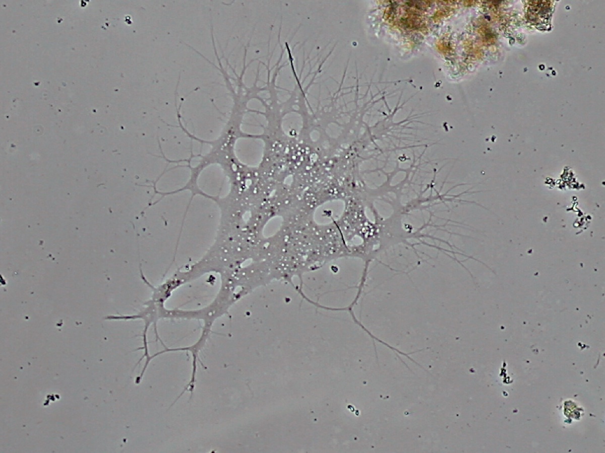
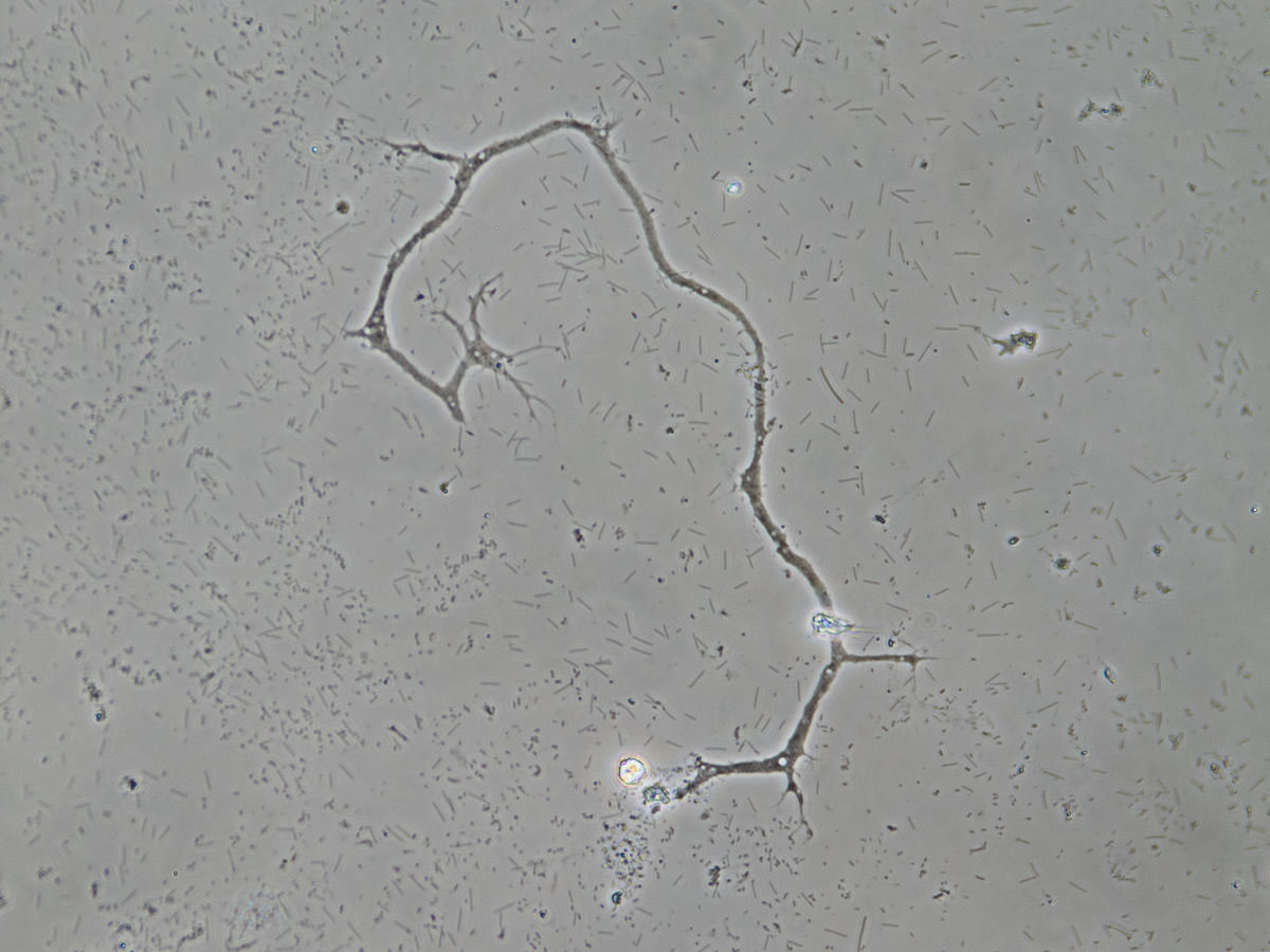
- Filopodia: fine filamentous pseudopodia, containing no granuloplasm or microtubules, sometimes branching but never anastomosing.
- Granulopodia: filamentous pseudopodia, with granules (extrusomes), sometimes branching and anastomosing.
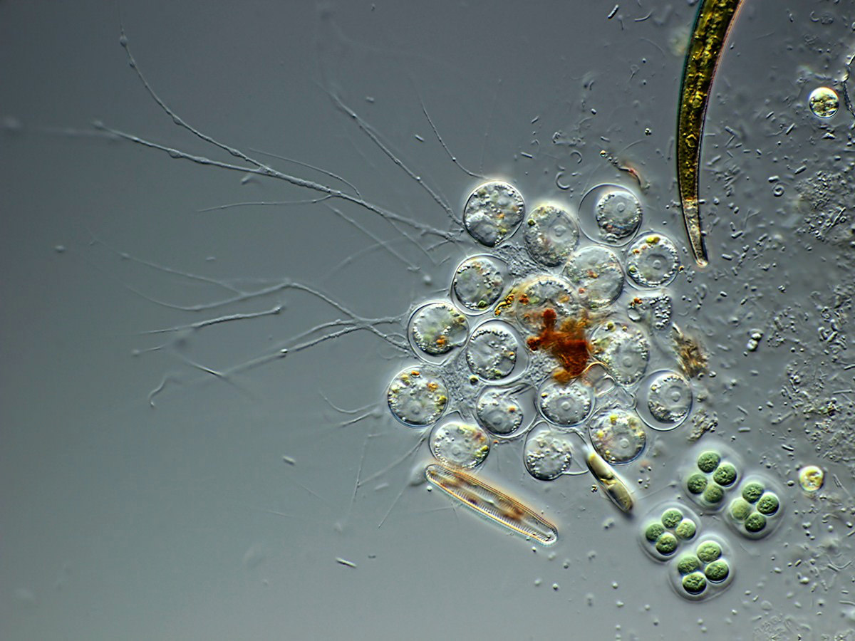
- Reticulopodia: fine filamentous pseudopodia without granules; often branching and anastomosing. Can form a network that is shared by multiple cells.
- Granuloreticulopodia: with granules, branching, anastomosing. Supported by microtubules.
Heliozoan Morphotype
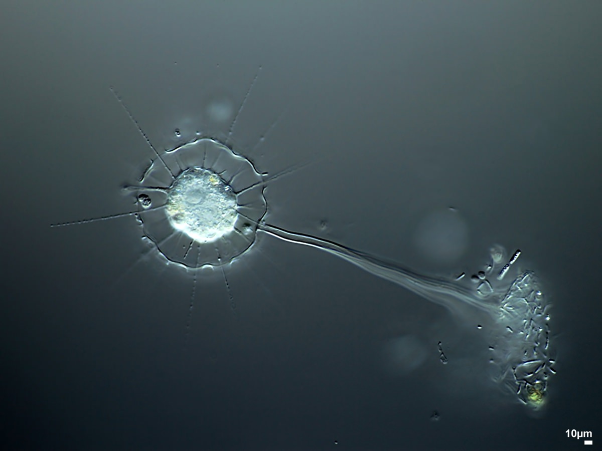
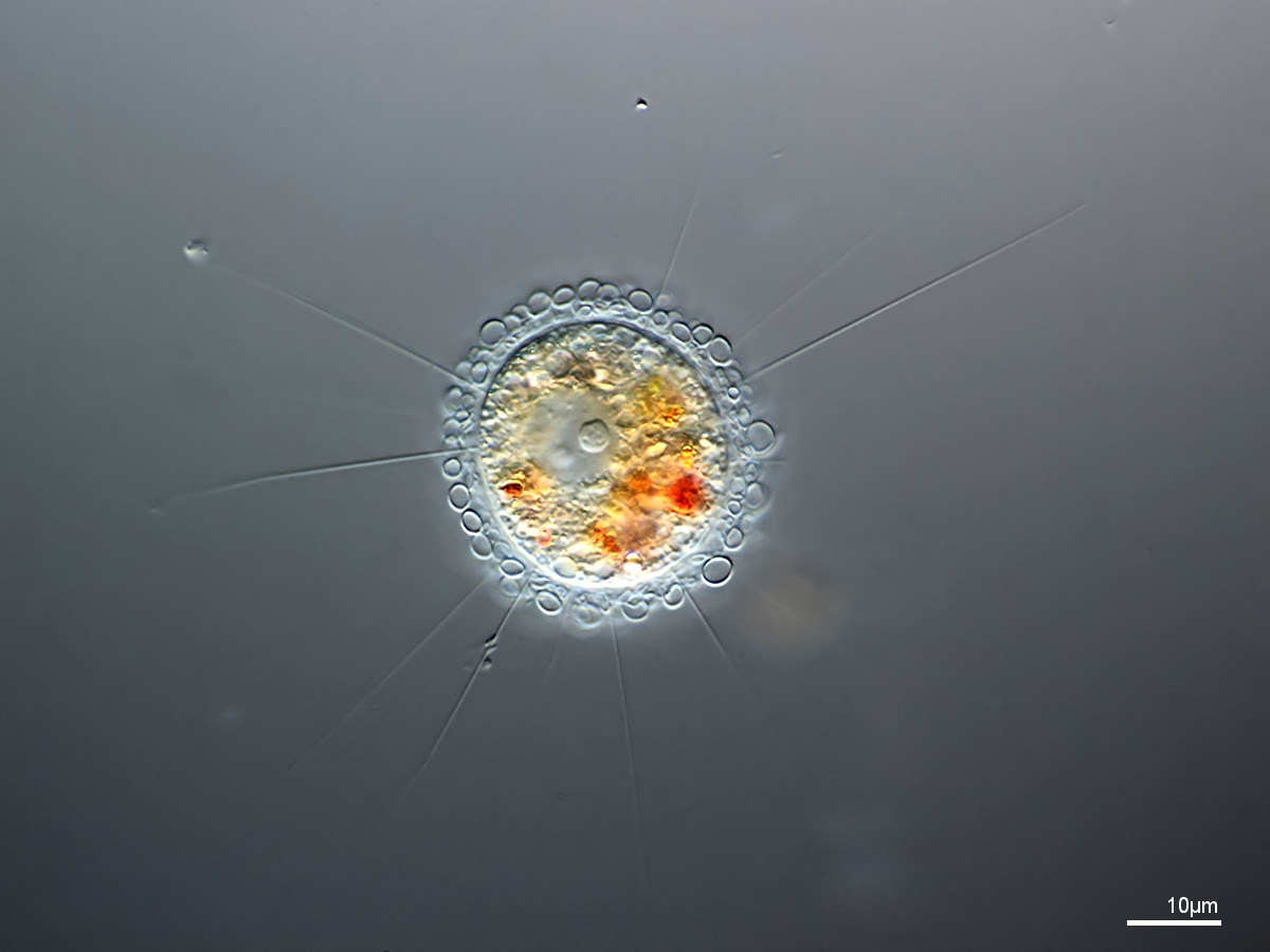
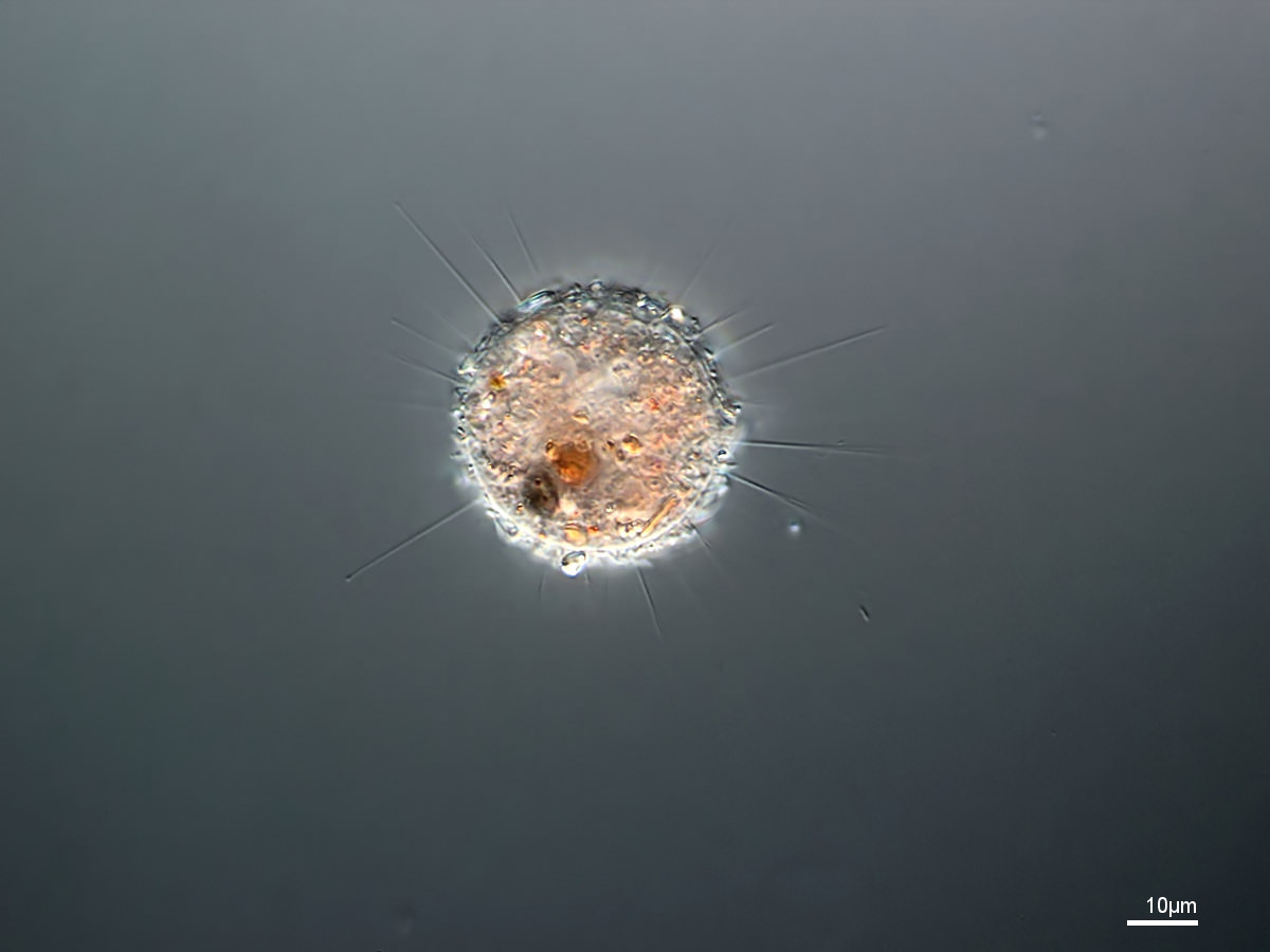
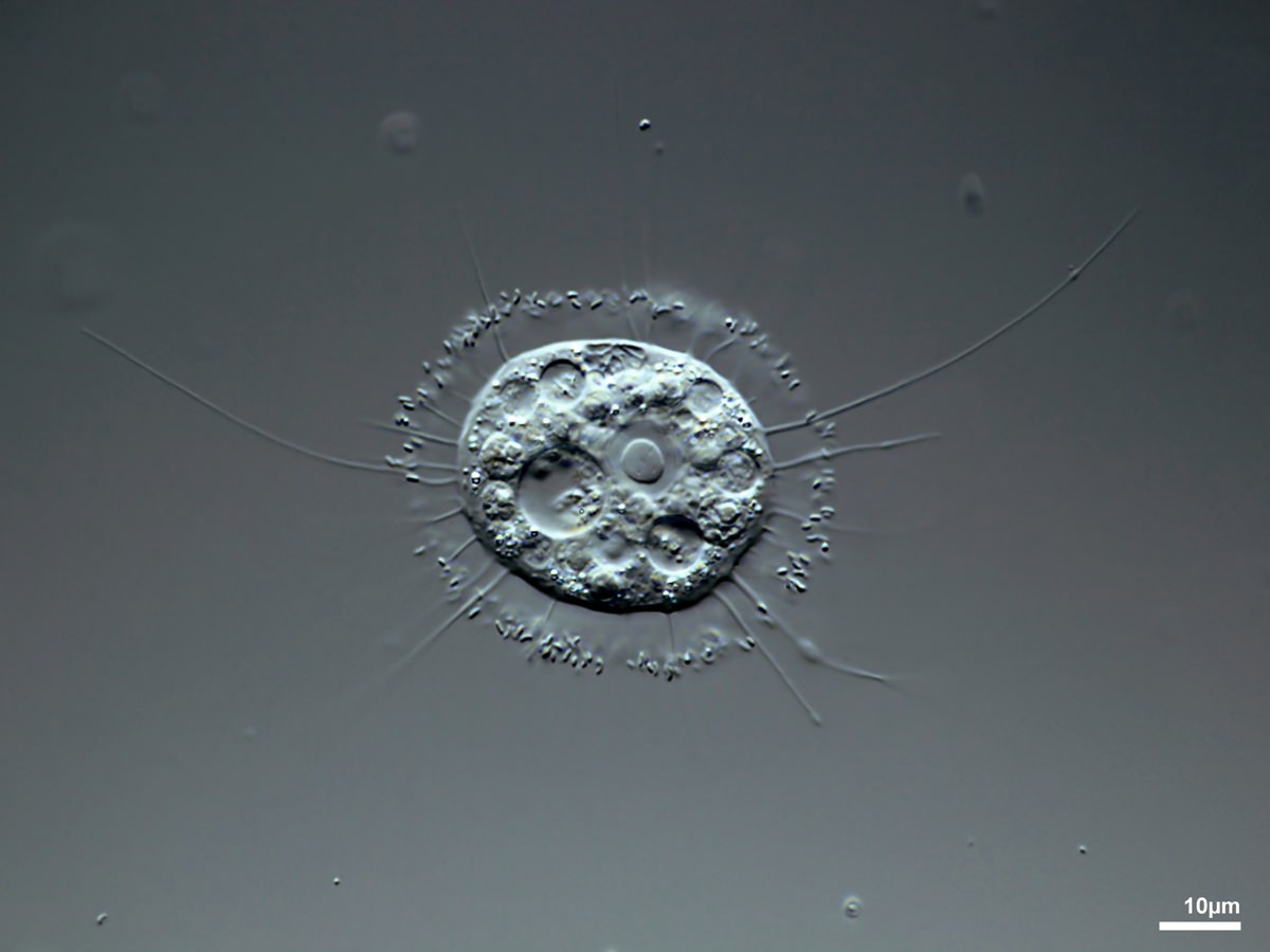
More or less spherical; floating in the water column, on the ground, or attached to a stalk. Often with spines and scales.
More or less spherical; floating in the water column, on the ground, or attached to a stalk. Often with spines and scales.
Pseudopodia: plasma projections with feeding, and/or sensing, and/or locomotion function.
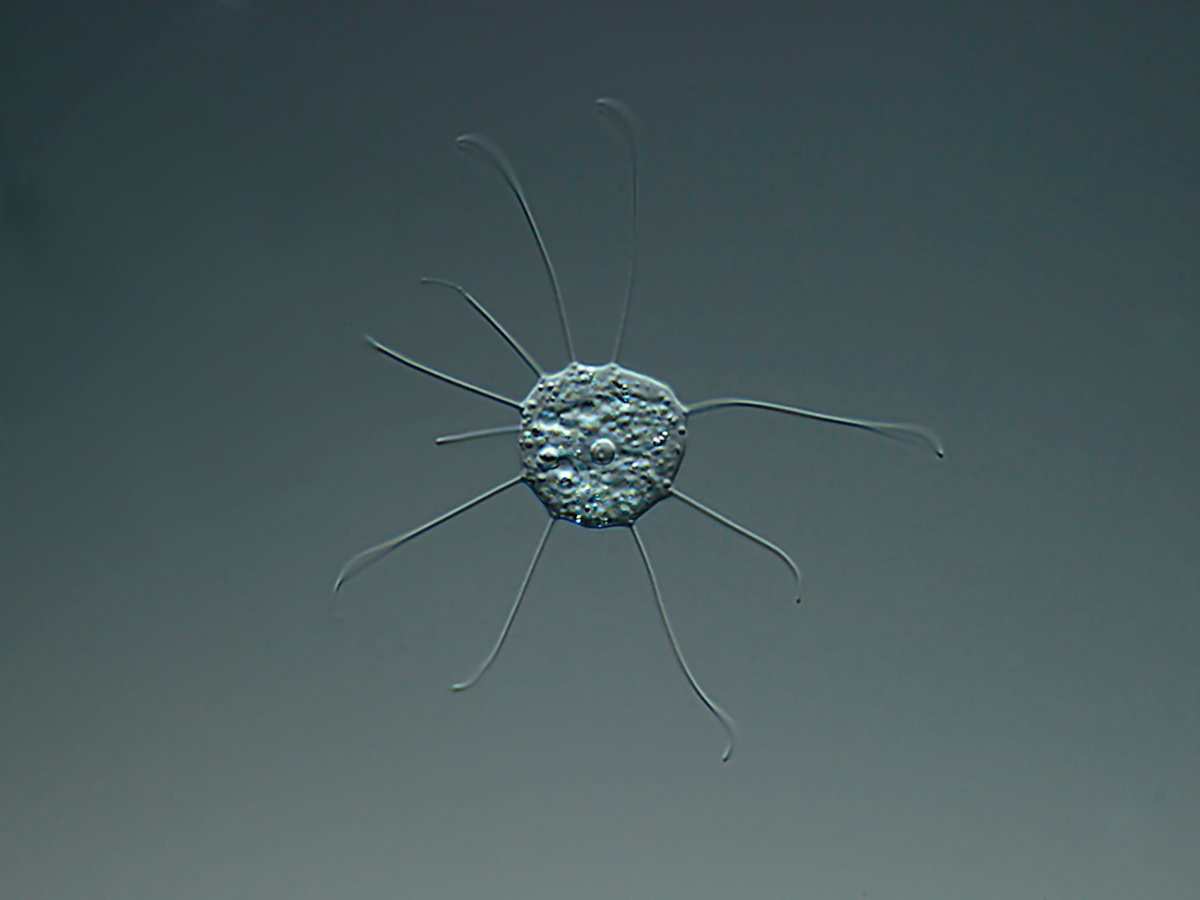
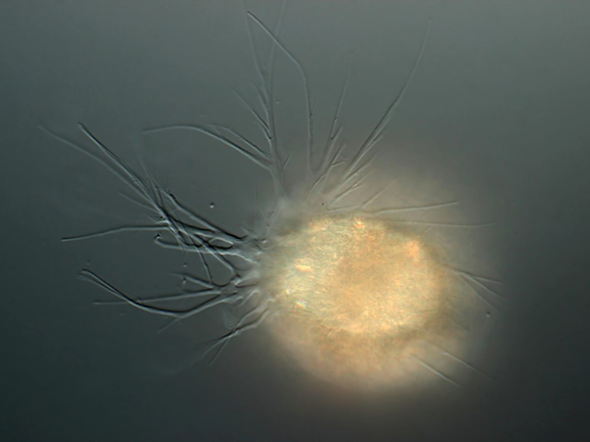
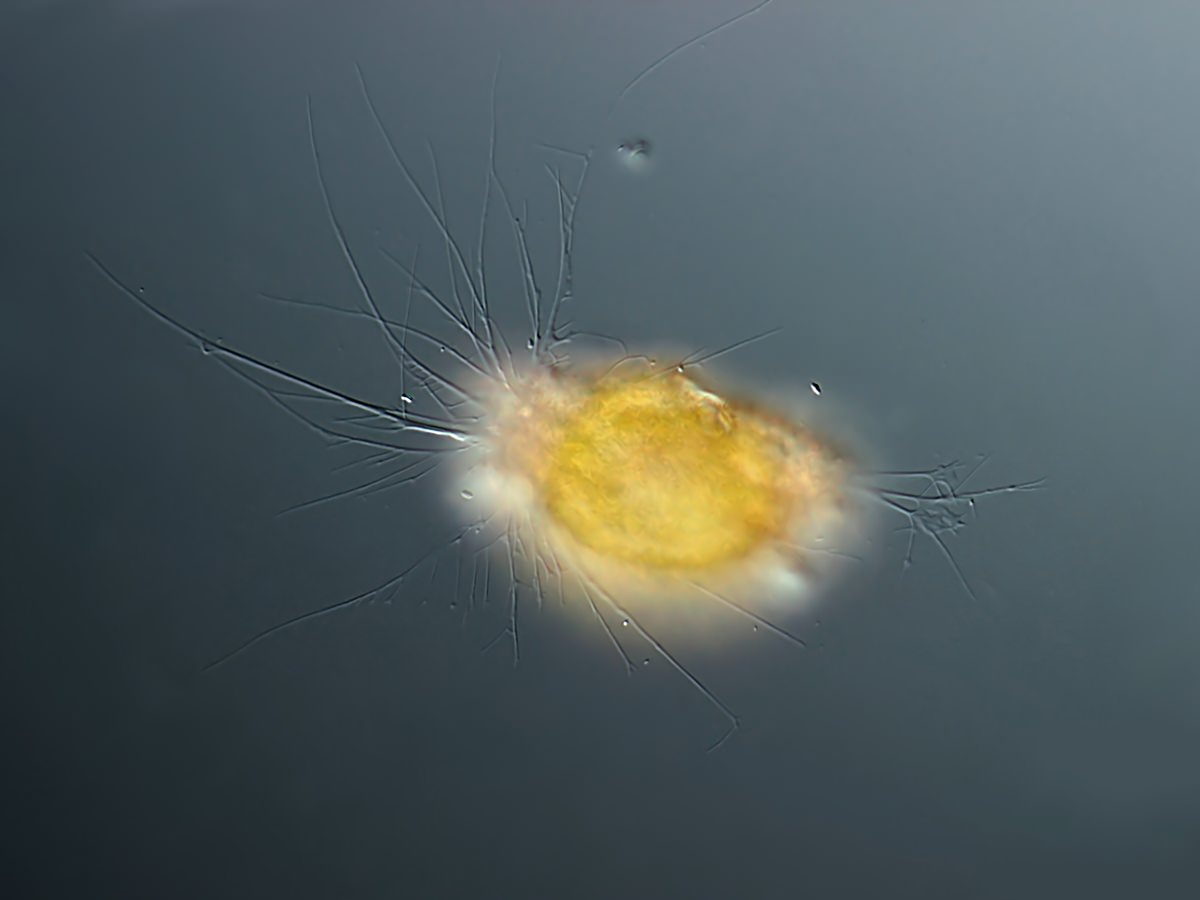
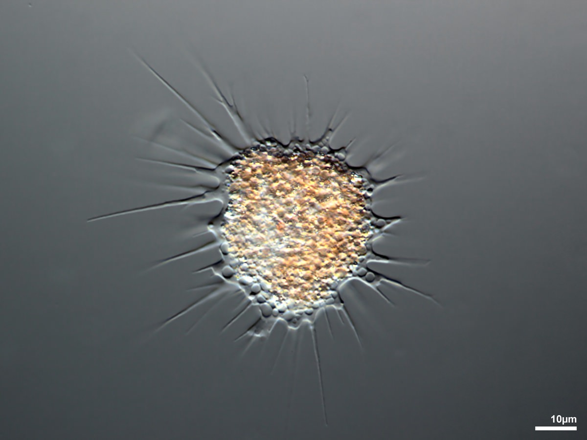
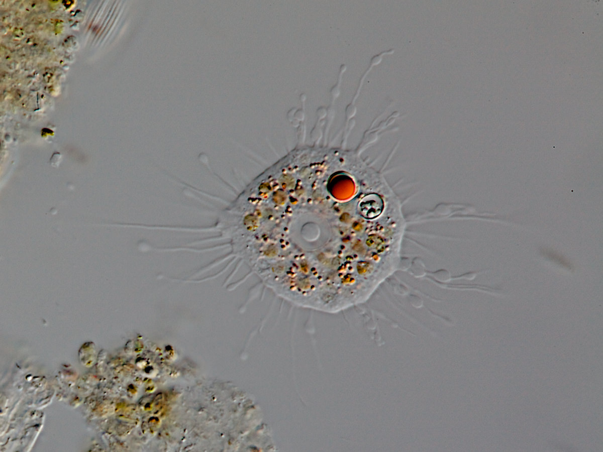
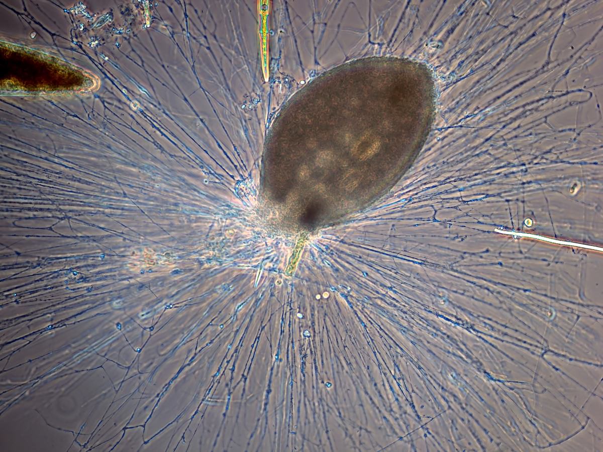
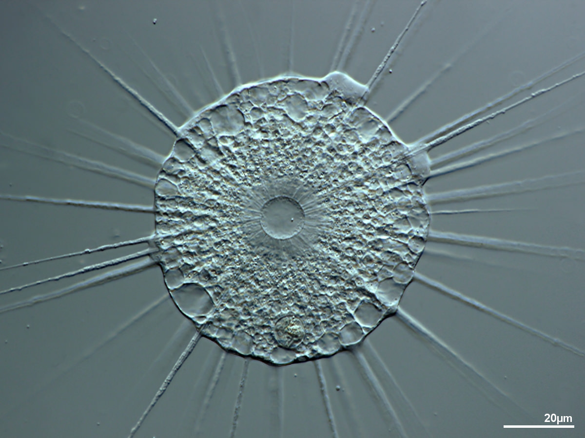
- Axopodia: ray-shaped pseudopodia with a stiff microtubullar core and extrusomes.
Axiopodia insert at a prominent, central nucleus
Axiopodia insert at multiple small nuclei
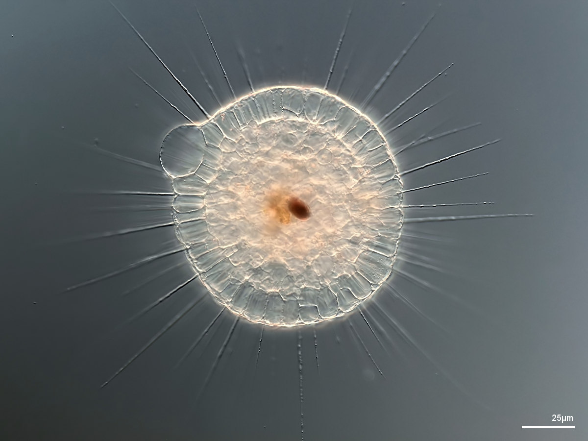
In the heliozoan form, axiopodia insert at the central nucleus, with a small cilium and a tiny, often invisible stalk. Axiopodia can retract and the cell can form a spindle-like shape with the cilium pointing forward.
Axiopodia insert at a centrosome, nucleus excentric. Often with scales, or scales and spicula.
Cell inside a chitinous shell that is perforated with numerous rather large openings, often stalked.
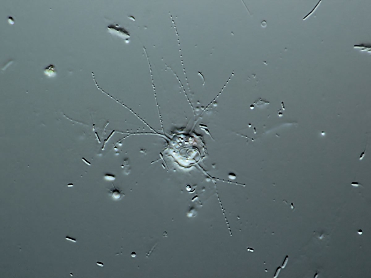
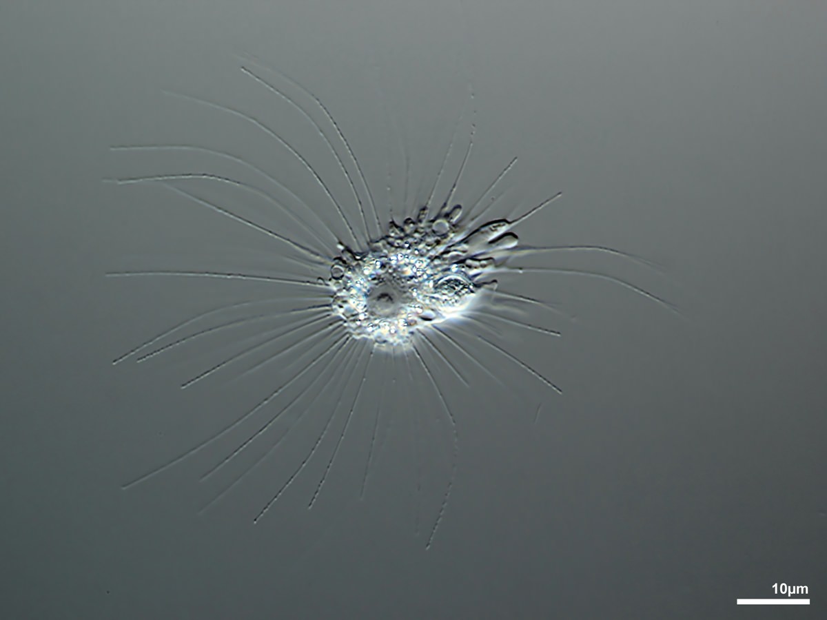
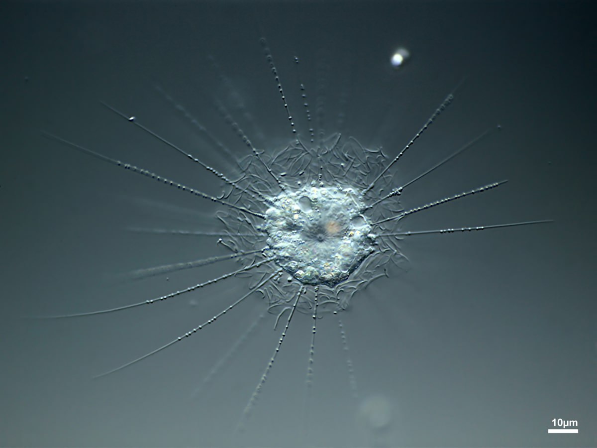
- Filopodia: ray-shaped thin pseudopodia without extrusomes and without a stiff microtubular core.
Protoplast covered with siliceous pearls or scales.
Protoplast covered with small sand particles.
Cell naked, often with a mucous coating and bacteria.
Cell covered by a thin test, multiple openings for filopodia, rapid movement by „waving“ filopodia.